STRIAE
DISTENSAE
MEDICAL MICRONEEDLING
An evidence-led approach to stretch-mark remodeling. Explore striae biology, clinical research, treatment planning and documented outcomes with medical mechanical microneedling.

Visible Striae Reduction
Less noticeable lines

Smoother Skin Texture
Improved surface quality

Better Skin Remodeling
More uniform appearance

Progressive Improvement
Results build gradually
Early striae at 6 months
Across randomized evidence
Documented by biopsy
Exceed striae study
Two clinical stages. One remodeling objective.
Striae rubrae
Red-to-violaceous striae represent the earlier, inflammatory stage. Assess both clinical appearance and lesion age before planning treatment: an evidence-supported acute-phase window is approximately 6 to <12 months. Lesions younger than six months should not simply be assumed to follow the same treatment course. [1]
Striae albae
Pale, atrophic striae represent the later stage of tissue change. Lesions present for more than one year can be approached as chronic striae, with the treatment goal centred on progressive improvement in skin structure, texture and scar quality rather than rapid visual correction. [1]
Why medical microneedling?
Medical microneedling creates controlled micro-injuries that stimulate wound healing, neocollagenesis, fibroblast activity and extracellular-matrix remodeling. Structural change develops over time. A clearer improvement signal can emerge from around Day 60 onward, so early post-treatment appearance should not be used as the final assessment of response. [1]

Medical Microneedling for Striae Distensae
Treatment techniques, needle depth, clinical endpoints and post-treatment care.
See the treatment in practice
Watch PD Dr. med. Matthias Aust demonstrate medical microneedling for abdominal stretch marks, explaining treatment settings, practical techniques and clinical endpoints.
How do you optimize the technique?
Explore needle depth adjustments, skin stretching, circular movements and device settings for treating striae.
What indicates the treatment endpoint?
See how visible tissue elevation and pinpoint bruising help identify when sufficient treatment has been achieved.
Why is proper aftercare essential?
Learn how post-treatment cleansing, skin care and crust prevention support the healing process.
Complete the form to watch the expert video.
A repeatable procedure and follow-up framework.
Screen before starting the series
Assess lesion age, current skin condition, recent microneedling and UV exposure before planning treatment.
Striae should be at least six months old for the treatment approach represented here. Lesions from approximately 6 to <12 months fall into the acute-phase window, while lesions present for more than one year can be considered chronic.
Important limitations
Do not begin a series through active inflammatory skin disease in the treatment area. Also consider recent microneedling and significant recent UV exposure when assessing whether the skin is ready to treat.
Microneedling for striae distensae
Plan treatment as a structured series, returning to the same striae and documenting progressive change. [1]
Plan treatment as a series rather than a single procedure. For body striae on the lower abdomen, hips and thighs, treatments can be spaced approximately one month apart, with up to four sessions across a 60–90-day treatment period. [1]
Repeatable sites and consistent follow-up
Mark the same striae before each session and use fixed reference points or an individual template to return to the same treatment sites. Follow changes in surface structure, barrier function, hydration, pigmentation, firmness and elasticity over time rather than relying on general visual impression alone.
Time-dependent remodeling
Judge treatment response across the full treatment series, not after the first session alone. [1]
As remodeling progresses, structural differences between striae and intact skin can narrow. Useful longitudinal markers include skin-line structure, surface smoothness and wrinkle-like relief. Relative differences across the Exceed study narrowed from 71% to 17% for anisotropy, 44% to 22% for smoothness and 52% to 30% for wrinkles over 90 days [1].
Allow time before judging the result
A clearer structural response can become apparent from approximately Day 60 onward. Keep photography, treatment-site positioning and assessment conditions consistent through later visits, and allow sufficient time for remodeling before evaluating the overall response to the series.
Assess structure and scar quality
Combine objective skin assessment with structured patient and clinician evaluation. [1]
Follow changes in pigmentation, relief, texture, thickness, elasticity and overall scar appearance, together with the patient’s perception of the treated area. Observer scores across the Exceed study improved from 39.50 to 24.67 and patient scores from 32.00 to 22.67, with pigmentation, relief and overall impression among the strongest areas of change.
Reassess after the active treatment series
Include a later follow-up rather than evaluating the result only at the final treatment visit. Compare the same documented lesions with baseline and assess whether texture, relief and overall scar quality continue to improve after the active treatment period.
Medical Microneedling: Clinical Insights, Techniques & Outcomes | PD Dr. Matthias Aust
From acne scars to stretch marks: patient selection, needle depth, treatment endpoints, safety, and realistic clinical expectations.
From clinical expertise to better treatment decisions.
Join PD Dr. med. Matthias Aust for an expert discussion on medical microneedling, combining clinical experience with practical treatment insights.
Why does the treatment mechanism matter?
Discover how mechanical microneedling differs from RF treatments and how controlled microinjury supports skin regeneration.
How do you know when to stop?
Explore needle depth, skin stretching, treatment techniques, and pinpoint bleeding as a clinical endpoint without overtreatment.
What really matters for safe, effective microneedling?
Learn why scientific evidence, device quality, professional training, and realistic patient expectations are essential for clinical success.
A reproducible workflow, from assessment to follow-up.
01 · Assess and stage
Distinguish rubrae from albae; record lesion age, body site, width, colour, relief and skin type. Review pregnancy, weight change, puberty, corticosteroid exposure, endocrine disease and ongoing stretch. Confirm suitability before planning treatment. [1]
02 · Map and document
Use fixed treatment and measurement points. Keep camera distance, lighting, framing, posture and tissue tension consistent. The EXCEED study used individual templates and contralateral healthy-skin references. Record time since first treatment as well as session number. [1]
03 · Plan preparation
The study used 30 minutes of topical anesthesia before treatment. Anesthetic selection, exposure area and duration must follow local requirements and patient-specific assessment. The published preparation is a research method, not a universal prescription. [1]
04 · Prepare the field
The investigators removed anesthetic with water-soaked gauze, then degreased and disinfected the area. Follow current device and antiseptic instructions, allow volatile antiseptics to dry, and use a new sterile single-use cartridge for every treatment. [1]
05 · Treat as a series
The study delivered up to four approximately monthly sessions, with many visits roughly 24–38 days apart. Select depth, passes and endpoints only within the current IFU and training. Document actual settings, anatomy and response at each visit. [1]
06 · Support recovery and reassess
Use permitted aftercare and clear instructions for cleansing, sun exposure and expected reactions. The study repeated photographs and probe measurements over time. Allow weeks for structural change; early redness or swelling is not the final result. [1]
Adding more context.
Long-term follow-up in light and dark skin
Twenty-five adults received one to three consecutive monthly treatments.
All participants were rated at least 50% improved after an average of 1.8 treatments; 28% were rated more than 75% improved. Reported effects were transient erythema, with no infections or dyspigmentation reported.
Scope of the evidence
An uncontrolled series using a microneedling device and blinded clinical ratings. No topical or intralesional adjuncts were used. These findings support the broader treatment signal.
Disk-microneedle pilot study
Sixteen Korean volunteers with rubrae or albae received three treatments four weeks apart.
Seven participants (43.8%) showed marked-to-excellent improvement. Six were highly satisfied and eight somewhat satisfied. Mild pain, erythema and spot bleeding were reported.
Study boundary
A small pilot using a disk microneedle system, with photographs, biopsies and satisfaction assessments.
Biopsy-confirmed dermal remodeling
Twenty-two women with striae received a single percutaneous collagen induction treatment.
At six months, clinical assessment showed improved skin texture and tightening. Biopsies demonstrated increased type I collagen and elastin, together with remodeling of the collagen-elastin matrix. No change in pigmentation was reported.
Scope of the evidence
A small uncontrolled clinical series with six-month histologic follow-up. The findings provide direct tissue-level evidence of dermal remodeling after mechanical needling, but there was no untreated or active control group.
Clinical and histologic improvement
Forty women with striae were assigned to needling therapy or microdermabrasion with sonophoresis. The needling group received three treatments at four-week intervals.
Needling produced significantly greater clinical improvement. Histologic assessment after treatment also showed increased collagen, fibroblast numbers and epidermal thickness.
Scope of the evidence
A comparative clinical and histopathological study using an active comparator. The findings support both visible improvement and tissue remodeling following mechanical needling.
Significant collagen and elastin increase
Twenty women with striae alba and Fitzpatrick skin types III–IV received five monthly treatments with microneedling and a non-ablative fractional laser on opposite abdominal sides.
Both modalities improved striae. Biopsies showed significant increases in collagen and elastic fibers after the third and fifth treatments, with no significant difference between microneedling and the laser treatment.
Scope of the evidence
A randomized split-abdomen study combining clinical assessment with skin biopsies. The histologic findings provide objective evidence of dermal remodeling after mechanical microneedling.
Significant reduction in striae size
Forty patients with striae alba received four monthly treatments, with similar striae randomized to mechanical microneedling or fractional CO₂ laser.
The cross-sectional size of the largest striae decreased significantly after microneedling. At six-month follow-up, outcomes did not differ significantly between microneedling and fractional CO₂ laser, and no clinically significant adverse effects were reported.
Scope of the evidence
A randomized clinical trial with follow-up extending to ten months. Objective striae measurements support a measurable treatment effect from mechanical microneedling rather than relying on photography alone.
Improvement continued after treatment
Twenty-nine participants with striae rubrae or striae albae received four monthly mechanical microneedling treatments, with site-matched untreated regions serving as controls.
At six months, Manchester Scar Scale scores improved by 48.9% for striae rubrae and 41.6% for striae albae. Treated regions also showed significantly greater reductions in striae length than untreated controls, with improvement continuing between three and six months after treatment.
Scope of the evidence
A prospective controlled study comparing treated areas with site-matched untreated skin. The continued improvement after the treatment series supports an ongoing remodeling response rather than only an immediate procedural effect.
Controlled injury requires controlled care.
Day 0 · Immediate response
Temporary redness, tenderness, mild pain and pinpoint bleeding can occur. Responses vary with area, technique and individual healing. These reactions were reported in the broader needling literature. [3,10]
Days 1–7 · Aftercare
Follow the current IFU and clinic protocol for cleansing, sun protection and permitted products. Possible reactions include dryness, roughness, tightness, itching, peeling, bruising, bleeding and crusting. [9]
Weeks · Remodeling
Surface calm does not mean collagen remodeling is complete. Continue comparable photographs and repeatable assessments. Avoid interpreting short-term swelling or hydration as a lasting treatment result. [1]
Day 60–90 · Reassessment
The EXCEED series showed clearer objective trends from approximately Day 60. Review skin texture, scar relief and patient-reported change together; individual results and timing vary. [1]
3 months and beyond
Reassess stage, ongoing causes, documented change and whether further treatment is justified. A series should be planned around healing and the current device instructions. [1]
Risks remain
Mechanical needling avoids intentional RF heating but does not fully eliminate infection, pigment change, scarring or inappropriate treatment risks. Patient selection, sterile single-use cartridges, technique and peri-procedure products matter. [9,10]
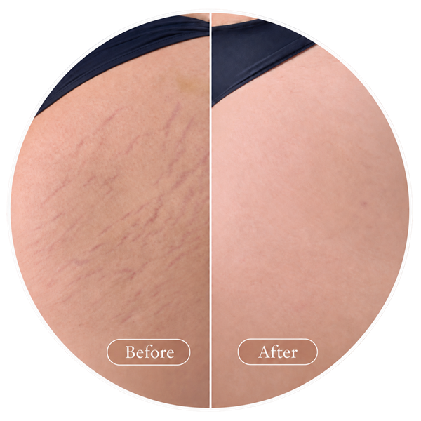
Document changes in stretch-mark appearance.


Image for illustrative purposes only. Courtesy of Dr. Zoya Evsyukova, specialist of regenerative medicine, plastic surgeon.
Texture, line pattern and surface appearance
Visible striae appear softer and less pronounced after treatment, with improved surface smoothness and reduced contrast against the surrounding skin.
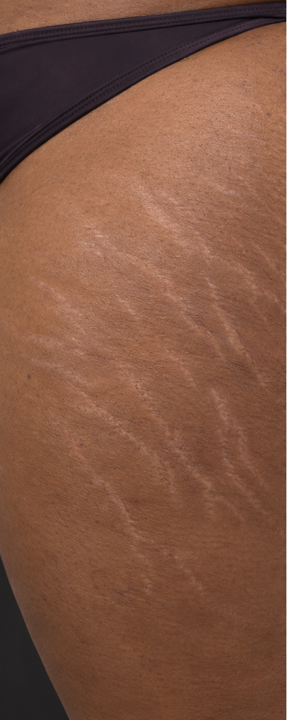
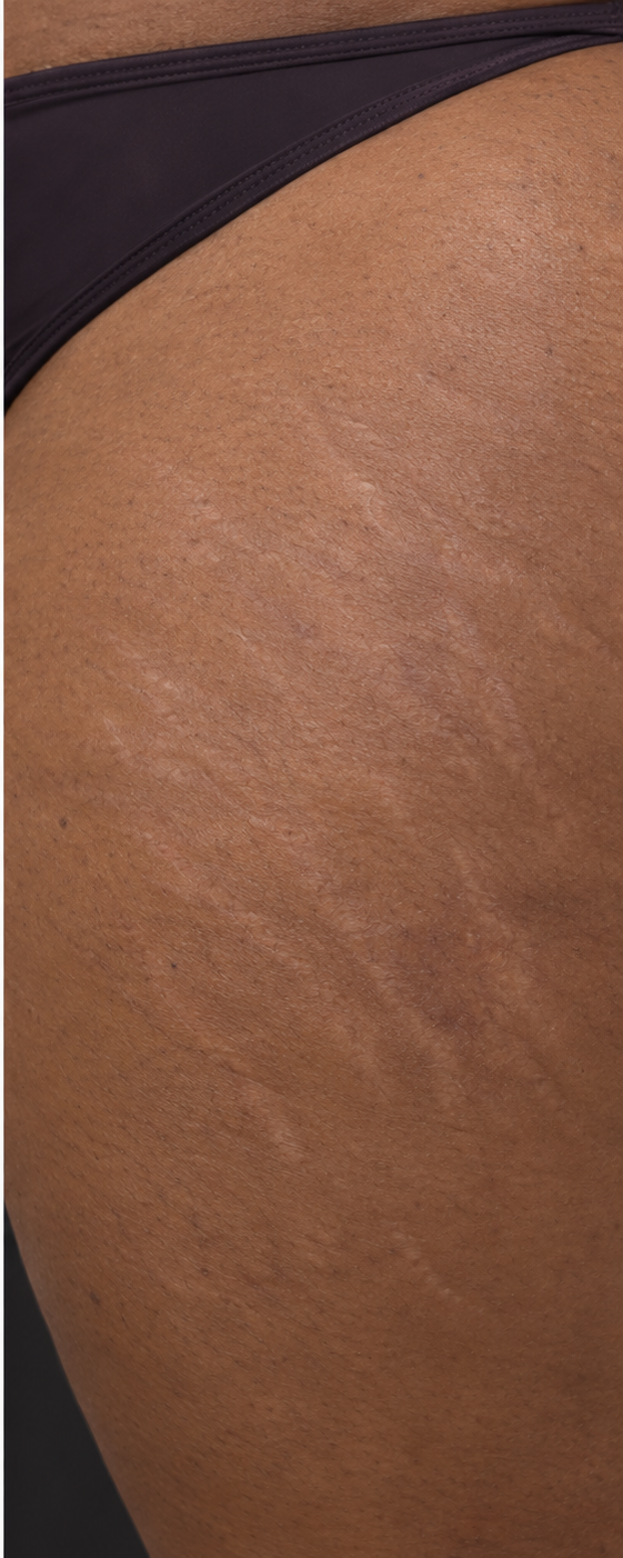
Image for illustrative purposes only. Courtesy of amiea med
Striae Albae in Dark Skin
Mature striae albae are clearly visible against darker skin. After treatment, contrast and textural irregularity decrease, while residual hypopigmented linear scars remain clinically recognizable.
Build a documented stretch-mark treatment pathway.
Explore medical microneedling
Review the evidence, consider stage and anatomy, and discuss training and product options for your market. Treatment should be performed by qualified professionals using the current regional labeling and Instructions for Use.
Set realistic expectations
The aim is measurable improvement over time. This educational page does not replace informed consent, clinical training or patient-specific medical judgment. Results vary, and striae indications differ by market.
Bring evidence into your clinical practice.
Discover EXCEED UNLIMITED and explore professional training, product information and options for your market. Review the evidence alongside the current regional labeling and Instructions for Use.
Scientific references
Medical microneedling for the treatment of striae distensae
[1] Kampmann AC, Bender R, Busch K, Bauer C, Hettwer K, Aust M. Medical microneedling for the treatment of striae distensae. A prospective study using the EXCEED Medical Needling Pen. Aesthetic Medicine. 2026;12(2):18363. DOI: 10.57662/AM.2026.18363.
Microneedling Treatment of Striae Distensae in Light and Dark Skin
[2] Alster TS, Li MK. Microneedling Treatment of Striae Distensae in Light and Dark Skin With Long-Term Follow-Up. Dermatol Surg. 2020;46(4):459–464.
Treatment of striae distensae using needling therapy
[3] Park KY, Kim HK, Kim SE, Kim BJ, Kim MN. Treatment of striae distensae using needling therapy: a pilot study. Dermatol Surg. 2012;38(11):1823–1828.
Comparison of the efficacy of microneedling versus CO₂ fractional laser
[4] Saki N, Rahimi F, Pezeshkian FS, Parvar SY. Comparison of the efficacy of microneedling versus CO₂ fractional laser to treat striae alba: a randomized clinical trial. Dermatol Ther. 2022;35(1):e15212.
Efficacy of fractional carbon dioxide laser versus microneedling
[5] Soliman M, Soliman MM, El-Tawdy A, Shorbagy HS. Efficacy of fractional carbon dioxide laser versus microneedling in the treatment of striae distensae. J Cosmet Laser Ther. 2019;21(5):270–277.
Microneedling Therapy for Striae Distensae
Sun X, Jia X, Huang L. Microneedling Therapy for Striae Distensae: Systematic Review and Meta-Analysis. Aesthetic Plast Surg. 2024;48(15):2915–2926.
Combined microneedling with topical PRP versus microneedling alone
[7] Abdel-Motaleb AA, Zedan H, Mostafa MM, et al. Combined microneedling with topical PRP versus microneedling alone in striae distensae: clinicopathological analysis. J Dermatolog Treat. 2022;33(2):836–847.
Microneedling: A Comprehensive Review
[8] Hou A, Cohen B, Haimovic A, Elbuluk N. Microneedling: A Comprehensive Review. Dermatol Surg. 2017;43(3):321–339.
U.S. Food and Drug Administration. Microneedling Devices
[9] U.S. Food and Drug Administration. Microneedling Devices: patient information and recommendations for health-care providers.
Safety Profile for Microneedling
[10] Safety Profile for Microneedling: A Systematic Review. PMID: 34448760.
Novel Therapeutic Option for Striae Distensae
[11] Aust MC, Knobloch K, Vogt PM. Percutaneous Collagen Induction Therapy as a Novel Therapeutic Option for Striae Distensae. Plast Reconstr Surg. 2010;126(4):219e–220e.
Needling Therapy Versus Microdermabrasion With Sonophoresis
[12] Nassar A, Ghomey S, El Gohary Y, El-Desoky F. Treatment of Striae Distensae With Needling Therapy Versus Microdermabrasion With Sonophoresis. J Cosmet Laser Ther. 2016;18(6):330–334.
Efficacy of Microneedling Versus Fractional Non-ablative Laser
[13] Naspolini AP, Boza JC, da Silva VD, Cestari TF. Efficacy of Microneedling Versus Fractional Non-ablative Laser to Treat Striae Alba: A Randomized Study. Am J Clin Dermatol. 2019;20(2):277–287.
Efficacy of Microneedling Versus CO₂ Fractional Laser
[14] Saki N, Rahimi F, Pezeshkian FS, Parvar SY. Comparison of the Efficacy of Microneedling Versus CO₂ Fractional Laser to Treat Striae Alba: A Randomized Clinical Trial. Dermatol Ther. 2022;35(1):e15212.
Safety and Efficacy of Microneedling
[15] Marin S, Watterson A, Alqam ML, Jones BC, Hitchcock TM. A Comparative Study to Evaluate the Safety and Efficacy of Microneedling as a Stand-Alone Treatment for Striae Rubrae and Albae. Aesthet Surg J. 2026;46(5):530–542.